Mammography Views and Positioning: A Guide
Precise mammography positioning is vital because it helps you get a full view of the anterior and posterior depth of the breast. Accurate mammo views significantly improve detection and diagnosis outcomes, helping patients receive early treatment when needed. With the right positioning, you can prevent artifacts, missed tissues and the need for repeated examinations while maintaining patient comfort.
Standard Mammography Views
Together, the following views provide standard imaging of the posterior and anterior depths of the breast:
The Craniocaudal (CC) View
The CC view shows medial to lateral tissue, distributing the tissue as much as possible for good radiation penetration. It provides a top-down image of the breast so you can visualize the tissue's posterior and inner or medial portions that may not appear in other views.
It's ideal to center the nipple and ensure that the posterior nipple line's (PNL's) length is within one centimeter of the PNL measured on the mediolateral oblique (MLO) view. You must position the image receptor beneath the breast and apply compression superiorly. With correct positioning, the CC view shows the deep medial breast tissue and detects masses that are typically excluded on the MLO view.
The Mediolateral Oblique (MLO) View
The MLO view offers a better view of the superior lateral quadrant, which is where the majority of the breast tissue is and where most breast cancers occur. This view shows tissue from the axilla to the inframammary fold, and it's important for capturing the most tissue, including the pectoralis muscle.
When positioning the breast for an MLO image, you must place the image receptor parallel to the pectoralis muscle to ensure the image includes maximum tissue and properly visualizes the axillary tail and axilla. The image receptor's angle may vary based on the patient's anatomy, so adjusting the gantry is essential for matching the muscle's angle. The compression paddle then compresses the breast superomedially. To ensure the proper acquisition technique, you must address three factors:
- The position of the pectoralis muscle: Ideally, the pectoralis muscle's margins should appear as convex curves or straight lines. This implies that you have sufficiently pulled the breast tissue forward.
- The PNL: The PNL and the pectoralis muscle should be able to intersect at a perpendicular angle.
- The inframmary fold (IMF): The IMF is the breast's most inferior portion and intersects the chest wall. Ensuring IMF inclusion in an MLO image helps you detect masses that may be located in the breast's lowest quadrant.
Advanced and Diagnostic Views
When standard mammography screening exams are inconclusive, the following specialized views can enhance diagnostic workups:
The Tangential View
The tangential view in mammography provides a clear view of a specific area of interest by projecting it away from other tissue. It's typically the best option for a palpable lump, but it can also help distinguish between intraparenchymal and dermal calcifications. With the tangential view, you must mark the area of interest and align the breast so that the space between the nipple and the marked area is tangential to the beam. Accurate positioning decreases superimposition of the tissue, ensuring you can clearly visualize the area of concern.
Exaggerated Craniocaudal (XCCL) View
The XCCL view is intended to visualize lateral breast tissue that a standard CC or MLO view might miss. This is especially useful for evaluating lateral breast masses or lesions near the chest wall. Rather than centering the breast, the XCCL view requires you to shift the imaging field laterally to capture more of the outer breast quadrants. To ensure accurate imaging with an XCCL view, you must do the following:
- Angle the C-arm laterally away from the midline while maintaining compression perpendicular to the chest wall.
- Ensure that the lateral breast tissue is fully stretched and positioned on the detector plate with the nipple in profile when possible.
- Avoid excessive compression that could obscure pathology.
- Include proper collimation in the lateral breast tissue and exclude any unnecessary chest wall.
- Verify that the pectoralis muscle does not obscure the breast's lateral tissue.
- Ensure the breast is adequately immobilized to prevent motion artifact.
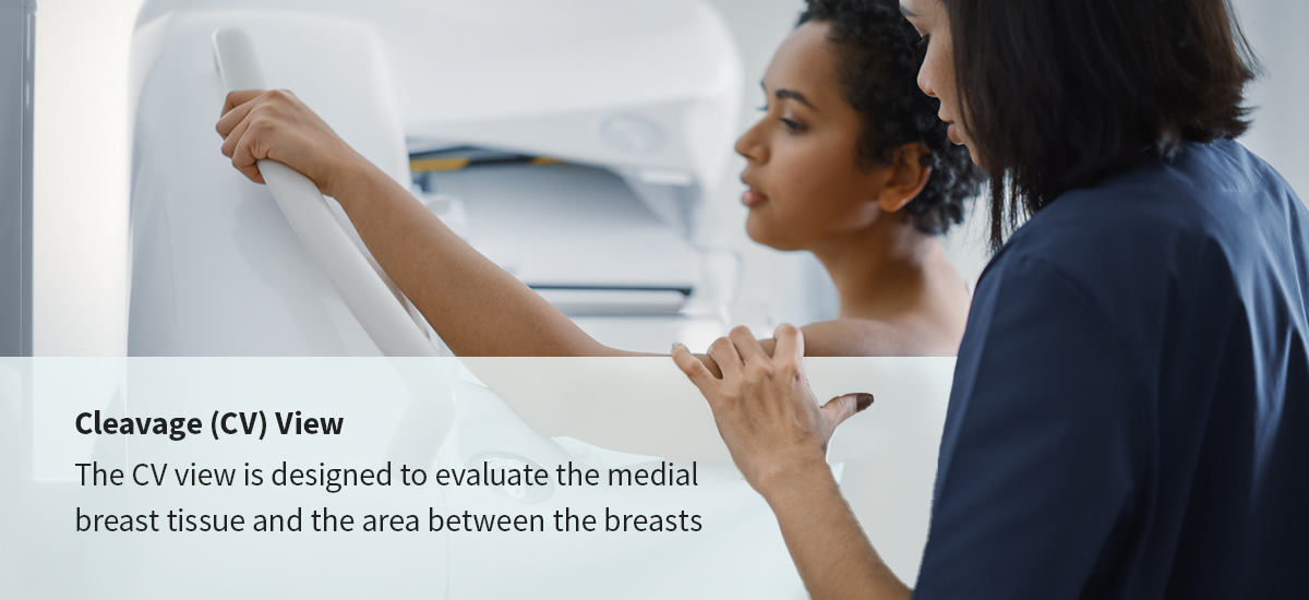
Cleavage (CV) View
The CV view is designed to evaluate the medial breast tissue and the area between the breasts, making it a valuable scan for evaluating the inframammary fold or detecting medial lesions. This view involves positioning both breasts on the detector simultaneously, which enables direct visualization of the cleavage area and medial aspects in a single image.
For proper positioning, the breasts' medial borders must touch or nearly touch at the midline. You must position the C-arm perpendicular to the detector and apply compression evenly and gently across both breasts. This helps maintain symmetry and prevents tissue distortion. You must also ensure the nipples are in profile, the inframammary fold is clearly visualized, and the medial tissue extends to the sternum without excessive pectoral muscle inclusion.
Implant-Displaced Views
Since implants appear dense on mammographic images and can obscure natural breast tissue, patients with breast augmentation should receive additional breast views apart from the standard screenings.
Standard CC and MLO views use lighter compression to include implants without rupturing them. Additional implant-displaced views require you to exclude as much of the implant as possible and perform the mammogram under normal compression. Implant-included views provide a general overview of the breast and a visualization of the implant, while implant-displaced views provide a closer evaluation of the natural breast tissue.
Proper MRI Breast Positioning
When a patient requires a diagnostic follow-up or a high-risk screening, breast MRI positioning is vital. While an MRI does not involve compression, it's important to ensure the patient remains immobile during the scan. Use immobilization pads, foam wedges and pillows to support the patient's body, and make sure the nipples are centered and pointing down before starting the scan.
From Positioning to Diagnosis: The Role of Technology
Even with perfect positioning, precise viewing and analysis software are necessary for accurate image interpretation and diagnosis. Upgrading to the following mammography equipment and software can significantly improve your ability to read, evaluate and share mammography images:
- Advanced mammography viewers: Candelis's Advanced Mammography Viewer is designed to enhance workflow in high-volume environments. It delivers faster read times, customizable hanging protocols and seamless integration with an enhanced Picture Archiving and Communication System (PACS).
- Efficient breast imaging software: Navigate complex 3D breast imaging datasets with fully integrated breast imaging software. Candelis's Breast Imaging Software can help you automate and streamline your workflows with reliable storage, cloud backup, automated image routing and easy prior study retrieval.
Enhance Your Workflow and Diagnostic Capabilities With Advanced Breast Imaging Solutions
Optimal mammography requires a combination of skilled positioning and advanced technical tools. Candelis provides advanced software solutions to empower your radiology department to achieve the highest standards of care. From image acquisition to final report, Candelis's Advanced Mammography Viewer and Breast Imaging Software can help you boost efficiency and improve diagnostics. Contact Candelis to learn more about breast imaging solutions and how they can improve your department's diagnostic capabilities and workflow.
- Log in to post comments